|
|
ECGbook.com Making Medical Education Free for All |
Upload ECG for Interpretation |
|
|
ECGbook.com Making Medical Education Free for All |
Upload ECG for Interpretation |
|
|
ECGbook.com Making Medical Education Free for All |


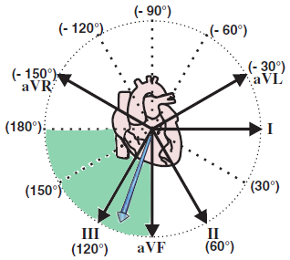
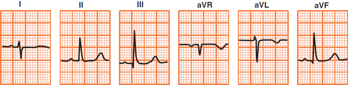
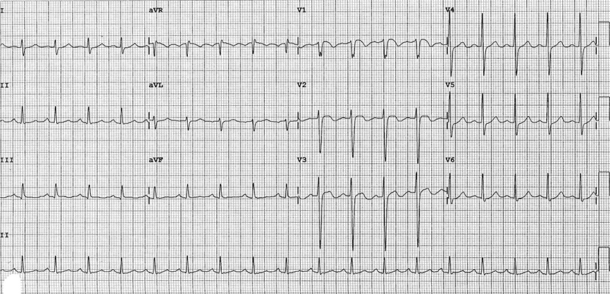
Right Axis Deviation

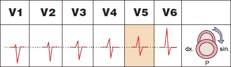
Clockwise Heart Rotation
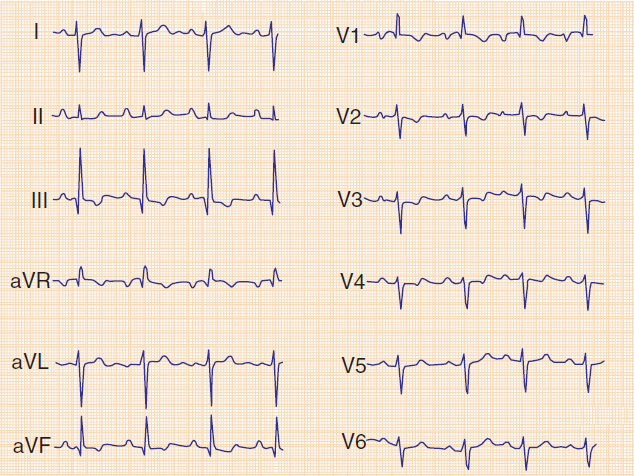
Acute Pulmonary Embolism
Acute Pulmonary Embolism
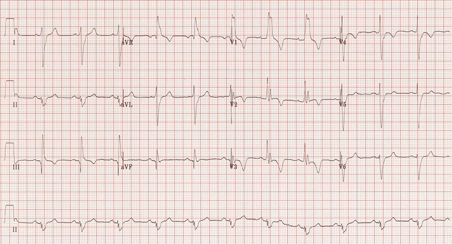
Acute Pulmonary Embolism
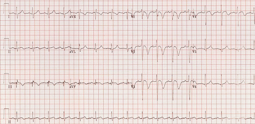
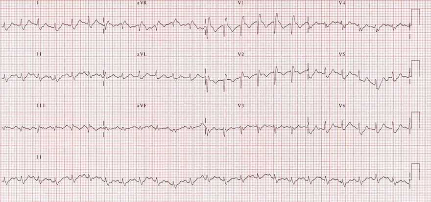
Massive Pulmonary Embolism
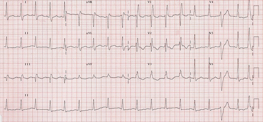
Acute Pulmonary Embolism
Sources
Cor Pulmonale
|
|
Pulmonary Embolism
|
|
|
|
|
|
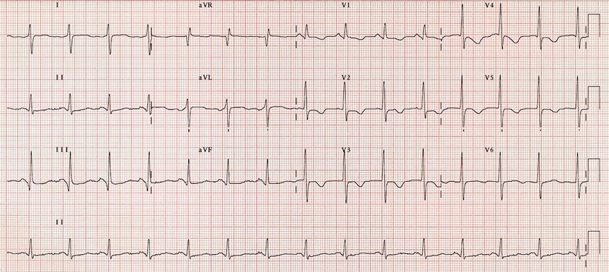
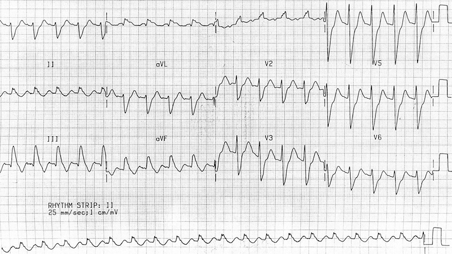
Right Axis Deviation
|
|
|
|
Clockwise Heart Rotation
Acute Pulmonary Embolism
Acute Pulmonary Embolism
Acute Pulmonary Embolism
Massive Pulmonary Embolism
Acute Pulmonary Embolism
Sources