|
|
ECGbook.com Making Medical Education Free for All |
Upload ECG for Interpretation |
|
|
ECGbook.com Making Medical Education Free for All |
Upload ECG for Interpretation |
|
|
ECGbook.com Making Medical Education Free for All |


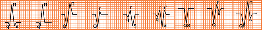
Nomenclature of the QRS Complex

Physiological Q Wave and Sinus Rhythm

Physiological Q Wave and Sinus Rhythm

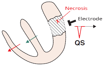
Pathological Q Wave and Old Infarction
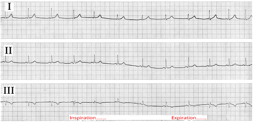
Positional (Respiratory) Q Wave
Sources
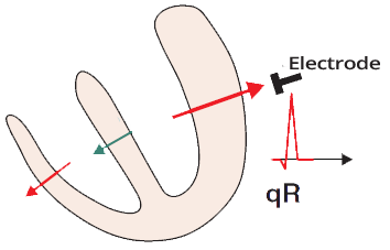
Mechanism of Q Wave Formation
|
|
Limb Leads and Q Wave
|
|
Precordial Leads and Q Wave
|
|
ECG and Q Wave
|
|
Pathological Q Wave
|
|
Nomenclature of the QRS Complex
Physiological Q Wave and Sinus Rhythm
Physiological Q Wave and Sinus Rhythm
Pathological Q Wave and Old Infarction
Positional (Respiratory) Q Wave
Sources