ECGbook.com

Making Medical Education Free for All

ECGbook.com

Making Medical Education Free for All

Home /

QRS Complex

QRS complex

Limb Leads and the QRS Complex

QRS complex morphology, septal vector, ventricular (main) heart vector, terminal vector in limb leads, vectorcardiogram
  • Limb leads (I, II, III, aVF, aVR, aVL)
    • View the heart in the frontal plane
  • If the direction of the electrical vector
    • Toward the lead - creates a positive deflection
    • Away from the lead - creates a negative deflection
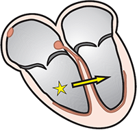
  • The ventricles are activated in the following order:
    1. Ventricular septum (VS - septal vector)
    2. Left and right ventricles (VM - main vector)
    3. Apex of the left ventricle (VT - terminal vector)
      • S wave
  • The three ventricular vectors create sequentially
    • the QRS complex

  • Each lead views the same vector from a different angle
    • The same vector appears differently in each lead

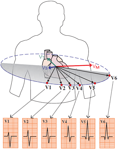
Precordial Leads and the QRS Complex

QRS complex morphology, septal vector, ventricular (main) heart vector, terminal vector in chest leads, vectorcardiogram

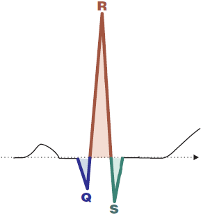
QRS Complex

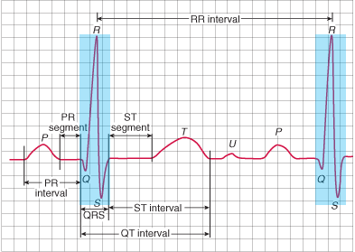
QRS complex morphology, Q wave, R wave, S wave
  • On an ECG, it shows ventricular depolarization
  • It is sometimes referred to as the QRS interval
  • Ventricular depolarization has 3 electrical vectors
    • Each vector has a different direction
  • Ventricular depolarization is always referred to as the QRS complex
    • Even if the QRS has a different configuration (RS, qR, Rs, R, rSR)
  • The ventricles have a more massive myocardium than the atria
    • Therefore, the P wave is smaller (atrial depolarization)
  • Ventricular depolarization (QRS complex) lasts 70ms - 110ms

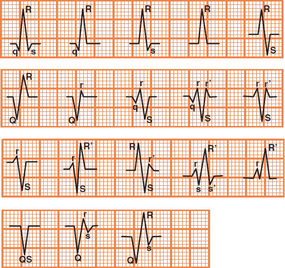
QRS Complex Nomenclature

QRS complex terminology, nomenclature, and correct interpretation

QRS Complex Nomenclature


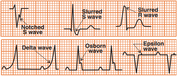
Fragmentation of the QRS Complex


QRS complex special cases, notched, slurred, delta Osborn, epsilon wave

Fragmentation of the QRS Complex


QRS Complex Concordance


ECG positive and negative QRS concordance, ventricular tachycardia

Ventricular Tachycardia


QRS Complex Width

  • Has a width of 70ms - 110ms (max. 2.5 squares)
  • The width of the QRS complex depends on the method of ventricular depolarization
    • Narrow QRS complex (< 110ms)
    • Wide QRS complex (>110ms)

Normal QRS complex width and duration


ECG narrow qrs complex (supraventricular origin), and broad qrs complex (ventricular origin)

Bigeminal Ventricular Extrasystoles

Narrow QRS Complex (<110ms)

narrow supraventricular QRS complex

ECG narrow supraventricular qrs complex - sinus rhythm

Sinus QRS Complexes


Wide QRS Complex (>110ms)

broad ventricular QRS complex

ECG broad qrs complex and ventricular tachycardia

Ventricular Tachycardia


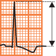
QRS Complex Amplitude

Increased QRS Complex Amplitude


ECG left ventricular hypertrophy, Sokolov Lyon voltage criteria (S wave depth in V1 + tallest R wave height in V5-V6 more than 35 mm)

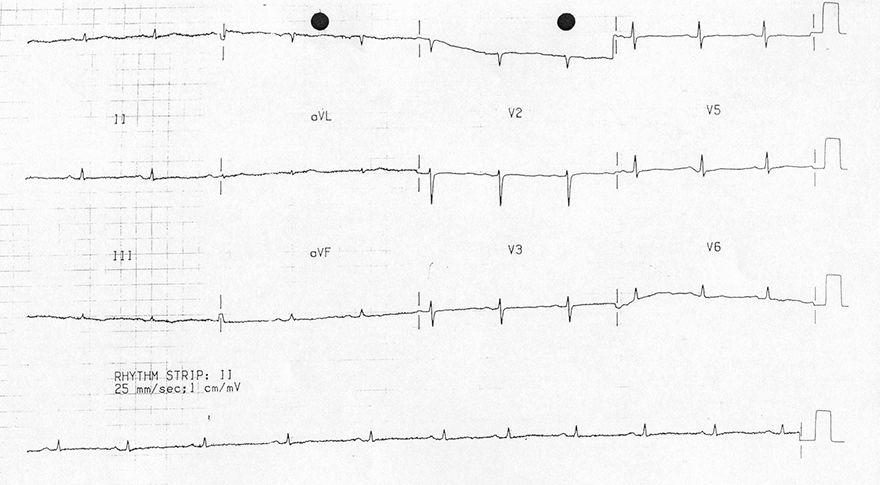
Decreased QRS Complex Amplitude


ECG low qrs voltage hypothyroidism, myxedema

Low QRS Voltage


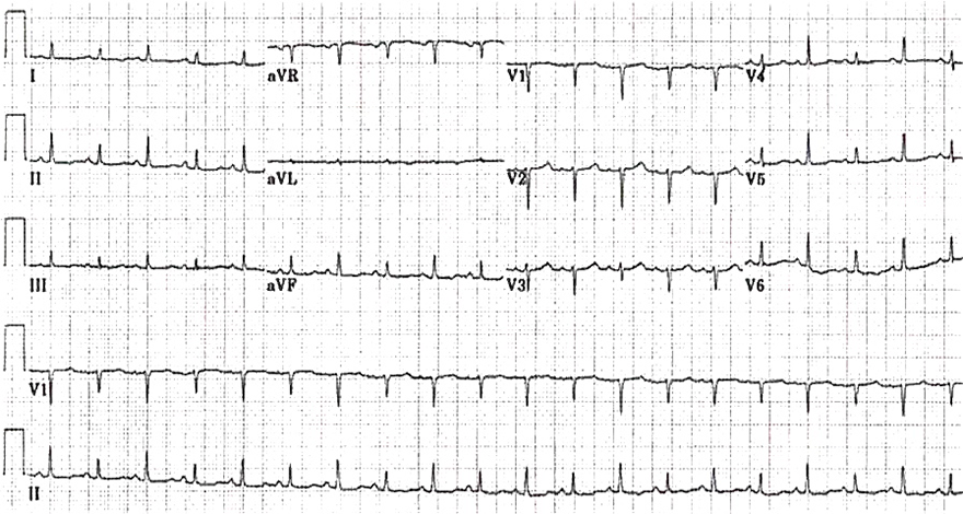
Electrical QRS Alternans


ECG QRS complexes alternate in height (voltage), massive pericardial effusion, heart swinging back and forth in pericardium

QRS Alternans


Sources

  • ECG from Basics to Essentials Step by Step
  • litfl.com
  • ecgwaves.com
  • metealpaslan.com
  • medmastery.com
  • uptodate.com
  • ecgpedia.org
  • wikipedia.org
  • Strong Medicine
  • Understanding Pacemakers


šípka späť

QRS Complex

QRS complex

Limb Leads and the QRS Complex

  • Limb leads (I, II, III, aVF, aVR, aVL)
    • View the heart in the frontal plane
  • If the direction of the electrical vector
    • Toward the lead - creates a positive deflection
    • Away from the lead - creates a negative deflection
  • The ventricles are activated in the following order:
    1. Ventricular septum (VS - septal vector)
    2. Left and right ventricles (VM - main vector)
    3. Apex of the left ventricle (VT - terminal vector)
      • S wave
  • The three ventricular vectors create sequentially
    • the QRS complex

  • Each lead views the same vector from a different angle
    • The same vector appears differently in each lead
QRS complex morphology, septal vector, ventricular (main) heart vector, terminal vector in limb leads, vectorcardiogram

Precordial Leads and the QRS Complex

QRS complex morphology, septal vector, ventricular (main) heart vector, terminal vector in chest leads, vectorcardiogram

QRS Complex

  • On an ECG, it shows ventricular depolarization
  • It is sometimes referred to as the QRS interval
  • Ventricular depolarization has 3 electrical vectors
    • Each vector has a different direction
  • Ventricular depolarization is always referred to as the QRS complex
    • Even if the QRS has a different configuration (RS, qR, Rs, R, rSR)
  • The ventricles have a more massive myocardium than the atria
    • Therefore, the P wave is smaller (atrial depolarization)
  • Ventricular depolarization (QRS complex) lasts 70ms - 110ms

QRS complex morphology, Q wave, R wave, S wave

QRS Complex Nomenclature

QRS complex terminology, nomenclature, and correct interpretation

QRS Complex Nomenclature


Fragmentation of the QRS Complex


QRS complex special cases, notched, slurred, delta Osborn, epsilon wave

Fragmentation of the QRS Complex


QRS Complex Concordance


ECG positive and negative QRS concordance, ventricular tachycardia

Ventricular Tachycardia


QRS Complex Width

  • Has a width of 70ms - 110ms (max. 2.5 squares)
  • The width of the QRS complex depends on the method of ventricular depolarization
    • Narrow QRS complex (< 110ms)
    • Wide QRS complex (>110ms)

Normal QRS complex width and duration


ECG narrow qrs complex (supraventricular origin), and broad qrs complex (ventricular origin)

Bigeminal Ventricular Extrasystoles


Narrow QRS Complex (<110ms)

narrow supraventricular QRS complex


ECG narrow supraventricular qrs complex - sinus rhythm

Sinus QRS Complexes


Wide QRS Complex (>110ms)

broad ventricular QRS complex


ECG broad qrs complex and ventricular tachycardia

Ventricular Tachycardia


QRS Complex Amplitude

Voltage QRS morphology, amplitude

Increased QRS Complex Amplitude

ECG left ventricular hypertrophy, Sokolov Lyon voltage criteria (S wave depth in V1 + tallest R wave height in V5-V6 more than 35 mm)

Decreased QRS Complex Amplitude


ECG low qrs voltage hypothyroidism, myxedema

Low QRS Voltage


Electrical QRS Alternans


ECG QRS complexes alternate in height (voltage), massive pericardial effusion, heart swinging back and forth in pericardium

QRS Alternans


Sources

  • ECG from Basics to Essentials Step by Step
  • litfl.com
  • ecgwaves.com
  • metealpaslan.com
  • medmastery.com
  • uptodate.com
  • ecgpedia.org
  • wikipedia.org
  • Strong Medicine
  • Understanding Pacemakers