|
|
ECGbook.com Making Medical Education Free for All |
|
|
ECGbook.com Making Medical Education Free for All |
|
|
ECGbook.com Making Medical Education Free for All |

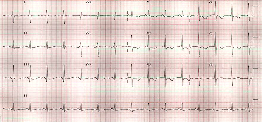
Normal (Intermediate) Heart Axis


Right Axis Deviation

Right Axis Deviation
Right Axis Deviation

Right Axis Deviation

Right Axis Deviation

Right Axis Deviation
Sources
Normal (Intermediate) Heart Axis
|
|
|
|
Right Axis Deviation
|
|
Right Axis Deviation
Right Axis Deviation
Right Axis Deviation
Right Axis Deviation
Right Axis Deviation
Sources