ECGbook.com

Making Medical Education Free for All

ECGbook.com

Making Medical Education Free for All

Home /

Right Ventricular Hypertrophy

Right ventricular hypertrophy

Right Ventricle

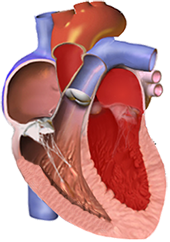
Normal right ventricle thickness
  • The right ventricle has a physiological thickness of 3-4mm
  • Hemodynamic overload of the ventricle (volume, pressure) causes remodeling of the ventricle
    • Right ventricular hypertrophy
      • It is thickening of the wall > 5mm
      • The wall of the right ventricle has a physiological thickness of 3-4mm
    • Left ventricular hypertrophy
      • It is thickening of the wall > 12mm
      • The wall of the left ventricle has a physiological thickness of 10-12mm
  • According to hemodynamic overload, we know 2 variants of hypertrophy:
    • Concentric hypertrophy
    • Eccentric hypertrophy

Concentric Hypertrophy of the Right Ventricle

right ventricle concentric hypertrophy

Eccentric Hypertrophy of the Right Ventricle

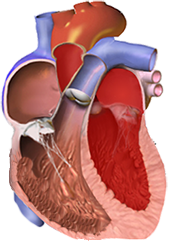
Eccentric right ventricular hypertrophy
  • The ventricle is overloaded with volume (increased preload)
    • In diastole, it is overfilled with blood
    • During systole, it pumps a larger volume of blood, also experiencing pressure overload
  • Dilation and hypertrophy of the ventricular cavity occurs
    • With chronic volume overload, only dilation occurs
  • Eccentric hypertrophy of the RV, causes:
    • Pulmonary insufficiency
  • Eccentric hypertrophy of the LV, causes:
    • Aortic insufficiency

Main Ventricular Vector and Ventricular Hypertrophy

ECG left ventricular hypertrophy, right ventricular hypertrophy, pattern features V1, V6

Right Ventricular Hypertrophy

  • ECG does not differentiate concentric hypertrophy from eccentric hypertrophy
  • The myocardium of the right ventricle (even with RV hypertrophy) is smaller than the myocardium of the left ventricle
    • The main cardiac vector, even with RV hypertrophy, does not point directly to the right ventricle
    • therefore, ECG changes in RV hypertrophy are minimal
  • ECG has only an orientational significance in the diagnosis of RV hypertrophy
  • Echocardiography is used for the diagnosis of hypertrophy

  • The principle of ECG diagnosis of RV hypertrophy is that the cardiac vector rotates:

ECG and Right Ventricular Hypertrophy


Dominant R Wave in V1

ECG right ventricular hypertrophy, dominant R wave in V1

Deep S Wave in V5/V6

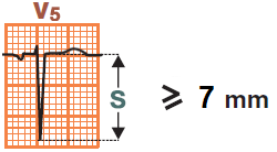
ECG right ventricular hypertrophy, dominant S wave in V5, V6


ECG right ventricular hypertrophy, voltage criteria, Right axis deviation, Dominant R wave in V1, Dominant S wave in V5 or V6

Right Ventricular Hypertrophy

  • Dominant R wave in V1 ≥ 7mm
  • Deep S wave in V5/V6 ≥ 7mm
  • Right axis deviation > +110°
    • Negative QRS in leads I and aVL
  • Right ventricular overload
    • Negative T waves in V1-2 without ST depressions

Right Ventricular Overload and Concentric RV Hypertrophy

Right ventricular strain pattern

ECG right ventricular strain pattern, right ventricular hypertrophy

Inferior Wall Infarction

  • Similar ECG changes to RV hypertrophy can be seen in inferior wall infarction (V1-3):
    • Dominant R wave in V1 ≥ 7mm (R/S ratio in V1 ≥ 1)
    • Horizontal ST depressions
    • Positive T waves (RV hypertrophy has negative T waves)
    • In inferior wall infarction, almost always there is an associated inferior wall infarction (ST elevations in II, III, aVF)
      • Because the posterior and inferior walls have a common blood supply through branches of the right coronary artery


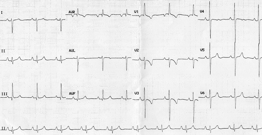
ECG right ventricular hypertrophy, Right axis deviation, Dominant R wave in V1, Dominant S wave in V6, Right ventricular strain pattern with ST depression and T-wave inversion in V1-4

Right Ventricular Hypertrophy


ECG right ventricular hypertrophy, Right axis deviation, Dominant R wave in V1, Dominant S wave in V6, Right ventricular strain pattern

Right Ventricular Hypertrophy

  • Dominant R wave in V1 ≥ 7mm
  • Deep S wave in V5/V6 ≥ 7mm
  • Right axis deviation > +110°
    • Negative QRS in leads I and aVL
  • Right ventricular overload (V1-3)
    • Descending ST depressions
    • Negative T waves


ECG right ventricular hypertrophy, P pulmonale, Right axis deviation, Dominant R wave in V1, Dominant S wave in V6, Right ventricular strain pattern

Right Ventricular Hypertrophy


ECG right vetricular hypertrophy, incomplete right bundle branch block (RBBB), Right ventricular strain pattern, Right axis deviation,

Right Ventricular Hypertrophy and Right Bundle Branch Block

  • Incomplete Right Bundle Branch Block:
    • rsR´ in V1
    • QRS duration < 0.12s
  • Incomplete RBBB with R´ in V1 ≥ 10 mm
  • Right axis deviation > +110°
    • Negative QRS in leads I and aVL
  • Right ventricular overload (V1-3) and (II, III, aVF)
    • Descending ST depressions
    • Negative T waves
  • P pulmonale
    • Height of P wave in II > 2.5mm


ECG right ventricular hypertrophy (RVH), arrhythmogenic right ventricular cardiomyopathy, dyplasia (ARVD), Right axis deviation, R S ratio

Right Ventricular Hypertrophy and Arrhythmogenic Right Ventricular Dysplasia


Sources

  • ECG from Basics to Essentials Step by Step
  • litfl.com
  • ecgwaves.com
  • metealpaslan.com
  • medmastery.com
  • uptodate.com
  • ecgpedia.org
  • wikipedia.org
  • Strong Medicine
  • Understanding Pacemakers


šípka späť

Right Ventricular Hypertrophy

Right ventricular hypertrophy

Right Ventricle

  • The right ventricle has a physiological thickness of 3-4mm
  • Hemodynamic overload of the ventricle (volume, pressure) causes remodeling of the ventricle
    • Right ventricular hypertrophy
      • It is thickening of the wall > 5mm
      • The wall of the right ventricle has a physiological thickness of 3-4mm
    • Left ventricular hypertrophy
      • It is thickening of the wall > 12mm
      • The wall of the left ventricle has a physiological thickness of 10-12mm
  • According to hemodynamic overload, we know 2 variants of hypertrophy:
    • Concentric hypertrophy
    • Eccentric hypertrophy


Normal right ventricle thickness

Concentric Hypertrophy of the Right Ventricle

  • The ventricle is overloaded with pressure (increased afterload)
    • The ventricle pumps blood against higher pressure
  • Hypertrophy occurs without dilation of the ventricular cavity
  • Concentric hypertrophy of the RV occurs with systolic overload of the RV:
  • Concentric hypertrophy of the LV occurs with systolic overload of the LV:
    • Arterial hypertension
    • Aortic stenosis


right ventricle concentric hypertrophy

Eccentric Hypertrophy of the Right Ventricle

  • The ventricle is overloaded with volume (increased preload)
    • In diastole, it is overfilled with blood
    • During systole, it pumps a larger volume of blood, also experiencing pressure overload
  • Dilation and hypertrophy of the ventricular cavity occurs
    • With chronic volume overload, only dilation occurs
  • Eccentric hypertrophy of the RV, causes:
    • Pulmonary insufficiency
  • Eccentric hypertrophy of the LV, causes:
    • Aortic insufficiency

Eccentric right ventricular hypertrophy

Main Ventricular Vector and Ventricular Hypertrophy

  • The main ventricular vector is best viewed in
  • Ventricular hypertrophy is seen on the ECG
    • Best in leads (V1 and V6)

  • Left ventricular hypertrophy (LVH)
    • The cardiac vector rotates towards LV hypertrophy
      • Because the LV is enlarged (LV hypertrophy)
    • The vector directs almost straight
  • Right ventricular hypertrophy (RVH)
    • The cardiac vector rotates towards RV hypertrophy
    • The vector is smaller, because the RV is smaller than the left
    • The vector directs almost straight
      • towards lead V1
      • away from lead V6
      • QRS amplitudes will be smaller
ECG left ventricular hypertrophy, right ventricular hypertrophy, pattern features V1, V6

Right Ventricular Hypertrophy

  • ECG does not differentiate concentric hypertrophy from eccentric hypertrophy
  • The myocardium of the right ventricle (even with RV hypertrophy) is smaller than the myocardium of the left ventricle
    • The main cardiac vector, even with RV hypertrophy, does not point directly to the right ventricle
    • therefore, ECG changes in RV hypertrophy are minimal
  • ECG has only an orientational significance in the diagnosis of RV hypertrophy
  • Echocardiography is used for the diagnosis of hypertrophy

  • The principle of ECG diagnosis of RV hypertrophy is that the cardiac vector rotates:

ECG and Right Ventricular Hypertrophy


Dominant R Wave in V1


ECG right ventricular hypertrophy, dominant R wave in V1

Deep S Wave in V5/V6

  • Deep S wave in V5/V6 ≥ 7mm

ECG right ventricular hypertrophy, dominant S wave in V5, V6


ECG right ventricular hypertrophy, voltage criteria, Right axis deviation, Dominant R wave in V1, Dominant S wave in V5 or V6

Right Ventricular Hypertrophy

  • Dominant R wave in V1 ≥ 7mm
  • Deep S wave in V5/V6 ≥ 7mm
  • Right axis deviation > +110°
    • Negative QRS in leads I and aVL
  • Right ventricular overload
    • Negative T waves in V1-2 without ST depressions

Right Ventricular Overload and Concentric RV Hypertrophy

Right ventricular strain pattern
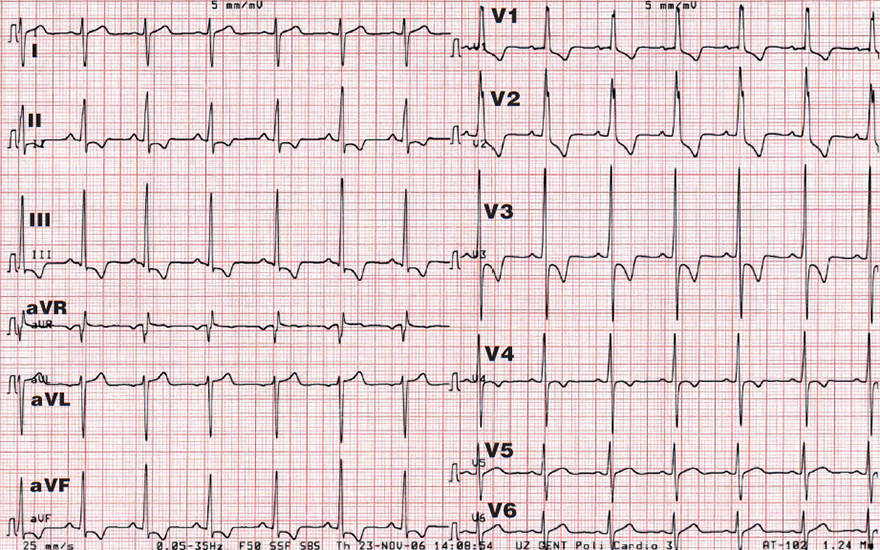
  • Caused by concentric RV hypertrophy (pressure overload)
    • Not by eccentric RV hypertrophy (volume overload)

  • In right-sided leads (V1-3), rarely in (II, III, aVF):
    • Descending ST depressions
    • Negative T waves

  • Occurs due to prolonged repolarization
ECG right ventricular strain pattern, right ventricular hypertrophy

Inferior Wall Infarction

  • Similar ECG changes to RV hypertrophy can be seen in inferior wall infarction (V1-3):
    • Dominant R wave in V1 ≥ 7mm (R/S ratio in V1 ≥ 1)
    • Horizontal ST depressions
    • Positive T waves (RV hypertrophy has negative T waves)
    • In inferior wall infarction, almost always there is an associated inferior wall infarction (ST elevations in II, III, aVF)
      • Because the posterior and inferior walls have a common blood supply through branches of the right coronary artery


ECG right ventricular hypertrophy, Right axis deviation, Dominant R wave in V1, Dominant S wave in V6, Right ventricular strain pattern with ST depression and T-wave inversion in V1-4

Right Ventricular Hypertrophy


ECG right ventricular hypertrophy, Right axis deviation, Dominant R wave in V1, Dominant S wave in V6, Right ventricular strain pattern

Right Ventricular Hypertrophy

  • Dominant R wave in V1 ≥ 7mm
  • Deep S wave in V5/V6 ≥ 7mm
  • Right axis deviation > +110°
    • Negative QRS in leads I and aVL
  • Right ventricular overload (V1-3)
    • Descending ST depressions
    • Negative T waves


ECG right ventricular hypertrophy, P pulmonale, Right axis deviation, Dominant R wave in V1, Dominant S wave in V6, Right ventricular strain pattern

Right Ventricular Hypertrophy


ECG right vetricular hypertrophy, incomplete right bundle branch block (RBBB), Right ventricular strain pattern, Right axis deviation,

Right Ventricular Hypertrophy and Right Bundle Branch Block

  • Incomplete Right Bundle Branch Block:
    • rsR´ in V1
    • QRS duration < 0.12s
  • Incomplete RBBB with R´ in V1 ≥ 10 mm
  • Right axis deviation > +110°
    • Negative QRS in leads I and aVL
  • Right ventricular overload (V1-3) and (II, III, aVF)
    • Descending ST depressions
    • Negative T waves
  • P pulmonale
    • Height of P wave in II > 2.5mm


ECG right ventricular hypertrophy (RVH), arrhythmogenic right ventricular cardiomyopathy, dyplasia (ARVD), Right axis deviation, R S ratio

Right Ventricular Hypertrophy and Arrhythmogenic Right Ventricular Dysplasia


Sources

  • ECG from Basics to Essentials Step by Step
  • litfl.com
  • ecgwaves.com
  • metealpaslan.com
  • medmastery.com
  • uptodate.com
  • ecgpedia.org
  • wikipedia.org
  • Strong Medicine
  • Understanding Pacemakers