ECGbook.com

Making Medical Education Free for All

ECGbook.com

Making Medical Education Free for All

Home /

Sinus Rhythm

Sinus rhythm, Normal sinus rhythm

Sinoatrial Node

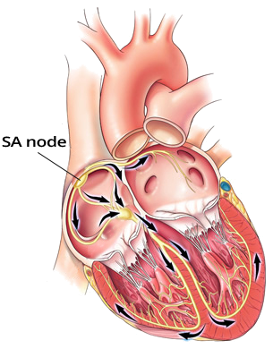
Pacemaker sinoatrial node (SA node) and conduction system
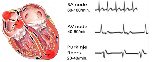
  • Automaticity
  • Overdrive Suppression
    • Impulses spread through the conduction system from the focus
      • which generates impulses at the highest frequency
    • A focus with the highest frequency deactivates foci
  • Sinoatrial Node (SA node)
    • Spontaneously generates impulses at a frequency of 60 - 100/min.
    • Other foci generate impulses with a lower frequency

Heart Rhythm

sinus rhythm, junctional rhythm, ventricular rhythm

Basic Heart Rhythms


P Wave and Sinus Rhythm

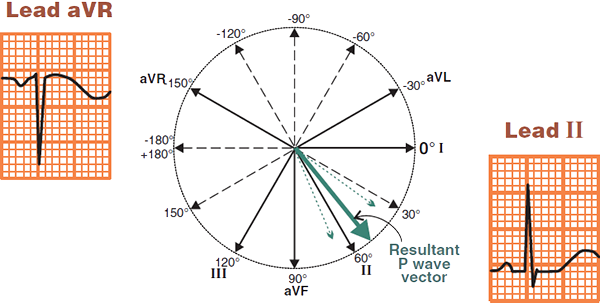
P wave electrical vector limb leads aVR, II

P Wave and Limb Leads

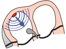
  • In sinus rhythm, impulses originate in the sinoatrial (SA) node
  • Each impulse propagates from the SA node towards the atria toward the AV node
    • This creates an atrial vector, which appears on the ECG as a P wave
  • The P wave is the primary characteristic of sinus rhythm
  • Physiological P wave
    • It is negative in lead aVR (because the vector is directed away from lead aVR)
    • It is positive in leads I, II (because the vector is directed towards leads I, II)

ECG and Sinus Rhythm

Sinoatrial SA node and sinus rhythm

ECG normal sinus rhythm

Sinus Rhythm


Frequency and Sinus Rhythm


ECG sinus tachycardia, electrical alternans

Sinus Tachycardia


ECG sinus bradycardia

Sinus Bradycardia


ECG sinus respiratory arrhythmia

Sinus Respiratory Arrhythmia

  • Average frequency: 90/min.
  • Heart rate is irregular (PP interval changes)
  • PQ interval is constant
  • P waves are present and are consistent, not indicative of Atrial Fibrillation
  • Sinus arrhythmia is a variant of normal, especially in young athletes, with SA node frequency changing:
    • During inhalation (sympathetic dominance) - SA node frequency increases
    • During exhalation (parasympathetic dominance) - SA node frequency decreases
  • It is a sinus rhythm (with respiratory arrhythmia), as impulses originate in the SA node


SA node sinus rhythm
ECG sinus rhythm, P wave morphology and axis (upright in I and II, inverted in aVR), Narrow QRS complexes, constant PR interval, regular rhythm

Sinus Rhythm


Sinoatrial node sinus rhythm
ECG sinus rhythm, narrow QRS, constant PQ PR interval, positive P wave II, negative P wave aVR

Sinus Rhythm

  • Frequency: 75/min.
  • Physiological P wave
    • Positive (I, II)
    • Negative (aVR)
  • Constant PQ interval (0.16s)
  • Narrow QRS complex (<0.12s)


Sinoatrial node sinus rhythm
ECG sinus rhythm, regular rhythm 75bpm, normal P waves, narrow QRS, constant PQ PR interval

Sinus Rhythm


sinoatrial node sinus rhythm
ECG respiratory sinus arrhytmia, irregular respiratory rhythm

Sinus Respiratory Arrhythmia

  • Average Frequency: 80/min.
  • Physiological P Wave
    • Positive (I, II)
    • Negative (aVR)
  • Heart Rhythm is Irregular (changes in PP interval and RR interval)
  • It is a sinus rhythm (with respiratory arrhythmia)
    • because impulses originate in the SA node
  • Constant PQ Interval (0.16s)
  • Narrow QRS Complex (<0.12s)


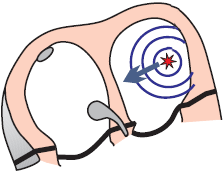
focal atrial tachycardia, ectopic atrial focus
ECG atrial focal tachycardia

Focal Atrial Tachycardia


atrial fibrillation, micro reentry
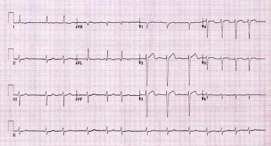
ECG irregular rhythm atrial fibrillation

Atrial Fibrillation

  • Frequency 80/min.
  • Heart rhythm is irregularly irregular (various RR intervals)
  • P waves are not visible, because impulses do not originate in the SA node
  • On the EKG, there is atrial fibrillation (the most common cardiac arrhythmia)
    • In the atria, there are multiple ectopic foci (micro re-entry)
    • Each focus generates its own vector and activates the atria
    • The atria have a frequency of 350-600/min., hence P waves are not visible
    • QRS complexes are irregularly irregular (various RR intervals)
      • Because impulses from the atria (350-600/min.)
        • are irregularly filtered through the AV node


Sources

  • ECG from Basics to Essentials Step by Step
  • litfl.com
  • ecgwaves.com
  • metealpaslan.com
  • medmastery.com
  • uptodate.com
  • ecgpedia.org
  • wikipedia.org
  • Strong Medicine
  • Understanding Pacemakers


šípka späť

Sinus Rhythm

Sinus rhythm, Normal sinus rhythm

Sinoatrial Node

  • Automaticity
  • Overdrive Suppression
    • Impulses spread through the conduction system from the focus
      • which generates impulses at the highest frequency
    • A focus with the highest frequency deactivates foci
  • Sinoatrial Node (SA node)
    • Spontaneously generates impulses at a frequency of 60 - 100/min.
    • Other foci generate impulses with a lower frequency

Pacemaker sinoatrial node (SA node) and conduction system

Heart Rhythm

sinus rhythm, junctional rhythm, ventricular rhythm

Basic Heart Rhythms


P Wave and Sinus Rhythm

P wave electrical vector limb leads aVR, II

P Wave and Limb Leads

  • In sinus rhythm, impulses originate in the sinoatrial (SA) node
  • Each impulse propagates from the SA node towards the atria toward the AV node
    • This creates an atrial vector, which appears on the ECG as a P wave
  • The P wave is the primary characteristic of sinus rhythm
  • Physiological P wave
    • It is negative in lead aVR (because the vector is directed away from lead aVR)
    • It is positive in leads I, II (because the vector is directed towards leads I, II)

ECG and Sinus Rhythm


Sinoatrial SA node and sinus rhythm


ECG normal sinus rhythm

Sinus Rhythm


Frequency and Sinus Rhythm


ECG sinus tachycardia, electrical alternans

Sinus Tachycardia


ECG sinus bradycardia

Sinus Bradycardia


ECG sinus respiratory arrhythmia

Sinus Respiratory Arrhythmia

  • Average frequency: 90/min.
  • Heart rate is irregular (PP interval changes)
  • PQ interval is constant
  • P waves are present and are consistent, not indicative of Atrial Fibrillation
  • Sinus arrhythmia is a variant of normal, especially in young athletes, with SA node frequency changing:
    • During inhalation (sympathetic dominance) - SA node frequency increases
    • During exhalation (parasympathetic dominance) - SA node frequency decreases
  • It is a sinus rhythm (with respiratory arrhythmia), as impulses originate in the SA node


ECG sinus rhythm, P wave morphology and axis (upright in I and II, inverted in aVR), Narrow QRS complexes, constant PR interval, regular rhythm

Sinus Rhythm

SA node sinus rhythm


ECG sinus rhythm, narrow QRS, constant PQ PR interval, positive P wave II, negative P wave aVR

Sinus Rhythm

  • Frequency: 75/min.
  • Physiological P wave
    • Positive (I, II)
    • Negative (aVR)
  • Constant PQ interval (0.16s)
  • Narrow QRS complex (<0.12s)
Sinoatrial node sinus rhythm


ECG sinus rhythm, regular rhythm 75bpm, normal P waves, narrow QRS, constant PQ PR interval

Sinus Rhythm

  • Frequency: 75/min.
  • Physiological P wave
    • Positive (I, II)
    • Negative (aVR)
  • Constant PQ interval (0.12s)
  • Narrow QRS complex (<0.12s)
Sinoatrial node sinus rhythm


ECG respiratory sinus arrhytmia, irregular respiratory rhythm

Sinus Respiratory Arrhythmia

  • Average Frequency: 80/min.
  • Physiological P Wave
    • Positive (I, II)
    • Negative (aVR)
  • Heart Rhythm is Irregular (changes in PP interval and RR interval)
  • It is a sinus rhythm (with respiratory arrhythmia)
    • because impulses originate in the SA node
  • Constant PQ Interval (0.16s)
  • Narrow QRS Complex (<0.12s)
sinoatrial node sinus rhythm


ECG atrial focal tachycardia

Focal Atrial Tachycardia

  • P wave has an abnormal shape
    • Negative (II, III, aVF)
    • Positive (V1)
    • Normal P wave is not negative (II, III, aVF) and positive (V1)
  • Frequency: 120/min.
  • This EKG does not show a sinus rhythm
focal atrial tachycardia, ectopic atrial focus


ECG irregular rhythm atrial fibrillation

Atrial Fibrillation

  • Frequency 80/min.
  • Heart rhythm is irregularly irregular (various RR intervals)
  • P waves are not visible, because impulses do not originate in the SA node
  • On the EKG, there is atrial fibrillation (the most common cardiac arrhythmia)
    • In the atria, there are multiple ectopic foci (micro re-entry)
    • Each focus generates its own vector and activates the atria
    • The atria have a frequency of 350-600/min., hence P waves are not visible
    • QRS complexes are irregularly irregular (various RR intervals)
      • Because impulses from the atria (350-600/min.)
        • are irregularly filtered through the AV node
atrial fibrillation, micro reentry


Sources

  • ECG from Basics to Essentials Step by Step
  • litfl.com
  • ecgwaves.com
  • metealpaslan.com
  • medmastery.com
  • uptodate.com
  • ecgpedia.org
  • wikipedia.org
  • Strong Medicine
  • Understanding Pacemakers