ECGbook.com

Making Medical Education Free for All

ECGbook.com

Making Medical Education Free for All

Home /

Sinus Tachycardia

Sinus tachycardia

Sinoatrial Node

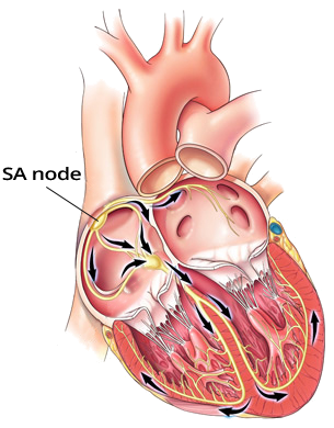
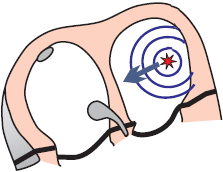
sinoatrial node (SA node) and heart conduction system
  • Automaticity
  • Overdrive Suppression
    • Impulses propagate through the conduction system from the focus
      • which generates impulses with the highest frequency
    • The focus with the highest frequency deactivates foci
  • Sinoatrial Node (SA Node)
    • Spontaneously generates impulses at a frequency of 60 - 100/min.
    • Other foci generate impulses at a lower frequency

Frequency and Sinus Rhythm


Sinus Tachycardia

  • Sinus Tachycardia is a sinus rhythm with a frequency >100/min.
    • In children, this means a frequency above the upper limit of normal
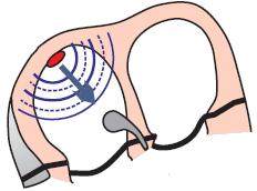
  • The SA node is intact, only generating impulses at a higher frequency (increased automaticity)
  • Sinus tachycardia is a secondary symptom - a sign of something else
    • The primary disease stimulates the SA node, increasing its frequency
    • In sinus tachycardia, one should always search for the primary cause
    • For example: a patient may primarily have angina with a fever, and secondarily (due to the fever) have sinus tachycardia

  • Maximum frequency of sinus tachycardia is 220 - age.
  • A 30-year-old patient has a maximum sinus tachycardia of 190/min. (220 - 30)
    • Sinus tachycardia rarely exceeds a frequency of 170/min.
  • Treatment of sinus tachycardia is causal, addressing the underlying cause of the sinus tachycardia (primary disease)

ECG and Sinus Tachycardia

SA node sinus tachycardia

ECG sinus tachycardia

Sinus Tachycardia


Most Common Causes of Sinus Tachycardia


Camel hump
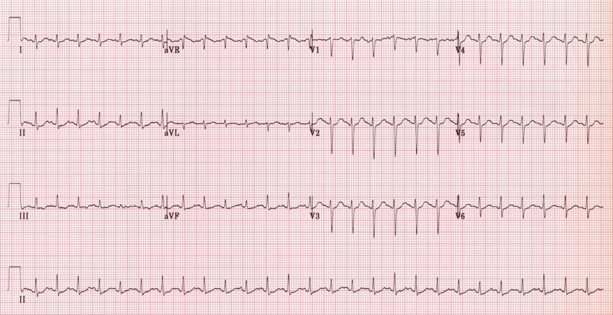
ECG camel hump appearance, hidden P waves in sinus tachycardia

Sinus Tachycardia

  • Frequency: 150/min. (>100/min.)
  • P waves are less visible because they overlap with T waves
  • P and T waves create two peaks, similar to the humps of a bactrian camel
    • This EKG pattern in sinus tachycardia is sometimes referred to as a "camel hump" in English


ECG normal sinus rhythm, heart rate 77 bpm

Sinus Rhythm


sa node sinus tachycardia
ECG sinus tachycardia, heart rate 150bpm, normal P waves, electrical qrs alternans

Sinus Tachycardia

  • Frequency: 150/min.
  • Physiological P wave
    • Positive (I, II)
    • Negative (aVR)
  • P waves merge closely with T waves and together form "two humps"
  • QRS alternans - changes in the amplitude of QRS complexes


sa node sinus tachycardia
ECG sinus tachycardia, heart rate 120bpm, normal P waves

Sinus Tachycardia


sa node sinus tachycardia
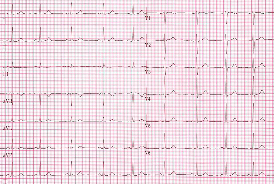
ECG sinus rhythm, heart rate 75bpm, normal P waves

Sinus Rhythm

  • Frequency: 75/min.
    • If the frequency were > 100/min., it would be sinus tachycardia
  • Physiological P wave
    • Positive (I, II)
    • Negative (aVR)


ectopic unifocal focus
ECG unifocal atrial tachycardia

Focal Atrial Tachycardia


Sources

  • ECG from Basics to Essentials Step by Step
  • litfl.com
  • ecgwaves.com
  • metealpaslan.com
  • medmastery.com
  • uptodate.com
  • ecgpedia.org
  • wikipedia.org
  • Strong Medicine
  • Understanding Pacemakers


šípka späť

Sinus Tachycardia

Sinus tachycardia

Sinoatrial Node

  • Automaticity
  • Overdrive Suppression
    • Impulses propagate through the conduction system from the focus
      • which generates impulses with the highest frequency
    • The focus with the highest frequency deactivates foci
  • Sinoatrial Node (SA Node)
    • Spontaneously generates impulses at a frequency of 60 - 100/min.
    • Other foci generate impulses at a lower frequency

sinoatrial node (SA node) and heart conduction system

Frequency and Sinus Rhythm


Sinus Tachycardia

  • Sinus Tachycardia is a sinus rhythm with a frequency >100/min.
    • In children, this means a frequency above the upper limit of normal
  • The SA node is intact, only generating impulses at a higher frequency (increased automaticity)
  • Sinus tachycardia is a secondary symptom - a sign of something else
    • The primary disease stimulates the SA node, increasing its frequency
    • In sinus tachycardia, one should always search for the primary cause
    • For example: a patient may primarily have angina with a fever, and secondarily (due to the fever) have sinus tachycardia

  • Maximum frequency of sinus tachycardia is 220 - age.
  • A 30-year-old patient has a maximum sinus tachycardia of 190/min. (220 - 30)
    • Sinus tachycardia rarely exceeds a frequency of 170/min.
  • Treatment of sinus tachycardia is causal, addressing the underlying cause of the sinus tachycardia (primary disease)

ECG and Sinus Tachycardia

SA node sinus tachycardia


ECG sinus tachycardia

Sinus Tachycardia


Most Common Causes of Sinus Tachycardia


ECG camel hump appearance, hidden P waves in sinus tachycardia Camel hump

Sinus Tachycardia

  • Frequency: 150/min. (>100/min.)
  • P waves are less visible because they overlap with T waves
  • P and T waves create two peaks, similar to the humps of a bactrian camel
    • This EKG pattern in sinus tachycardia is sometimes referred to as a "camel hump" in English


ECG normal sinus rhythm, heart rate 77 bpm

Sinus Rhythm


ECG sinus tachycardia, heart rate 150bpm, normal P waves, electrical qrs alternans

Sinus Tachycardia

  • Frequency: 150/min.
  • Physiological P wave
    • Positive (I, II)
    • Negative (aVR)
  • P waves merge closely with T waves and together form "two humps"
  • QRS alternans - changes in the amplitude of QRS complexes
sa node sinus tachycardia


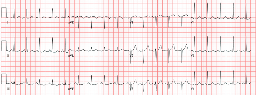
ECG sinus tachycardia, heart rate 120bpm, normal P waves

Sinus Tachycardia

  • Frequency: 120/min.
  • Physiological P wave
    • Positive (I, II)
    • Negative (aVR)
  • P waves are better assessed, do not merge with T waves
sa node sinus tachycardia


ECG sinus rhythm, heart rate 75bpm, normal P waves

Sinus Rhythm

  • Frequency: 75/min.
    • If the frequency were > 100/min., it would be sinus tachycardia
  • Physiological P wave
    • Positive (I, II)
    • Negative (aVR)
sa node sinus tachycardia


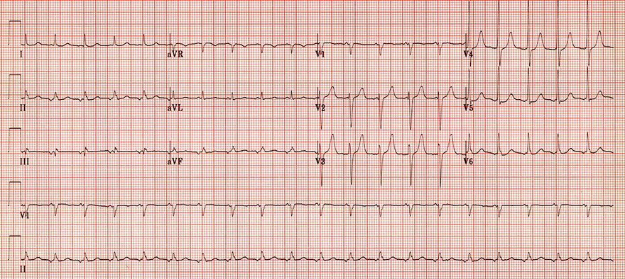
ECG unifocal atrial tachycardia

Focal Atrial Tachycardia

  • Frequency: 120/min.
  • P wave does not have a physiological shape
    • Negative (II, III, aVF)
    • Positive (V1)
    • Physiological P wave is not negative (II, III, aVF), and positive (V1)
  • This EKG does not show sinus rhythm
ectopic unifocal focus


Sources

  • ECG from Basics to Essentials Step by Step
  • litfl.com
  • ecgwaves.com
  • metealpaslan.com
  • medmastery.com
  • uptodate.com
  • ecgpedia.org
  • wikipedia.org
  • Strong Medicine
  • Understanding Pacemakers