ECGbook.com

Making Medical Education Free for All

ECGbook.com

Making Medical Education Free for All

Home /

STEMI and Ventricular Pacing

STEMI infarction and ventricular paced rhythm (Sgarbossa criteria)

Ventricular Pacing

paced ventricular rhythm and acute STEMI infarction
ECG ventricular paced rhythm with LBBB pattern
  • It is the ability of a pacemaker to stimulate the ventricular myocardium through an electrode
    • The electrode stimulates the myocardium with an electrical impulse (similar to the SA node)
  • The ventricular electrode of the pacemaker is placed in the right ventricle
  • Pacing on the ECG is seen as a pacing artifact (a vertical line, also known as a spike)
  • Ventricular pacing creates a LBBB pattern on the ECG
    • And LBBB masks the ECG signs of infarction

Sgarbossa Criteria


ECG and Acute STEMI with Ventricular Pacing

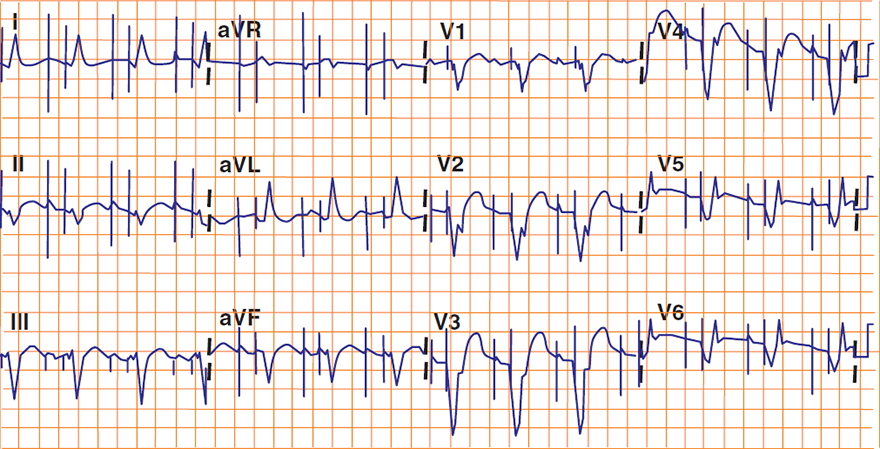
ECG Sgarbossa criteria STEMI infarction with ventricular paced rhythm, ST elevation, concordant ST depression
  • 1. Concordant ST elevation > 1mm (V4-V6, aVL, I)
    • In leads with a positive QRS complex
    • 5 points
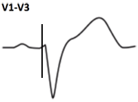
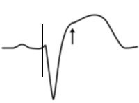
  • 2. Disconcordant ST elevation ≥ 5mm (V1-V3)
    • Or ST elevation > 25% of the depth of the S wave
    • In leads with a negative QRS complex
    • 2 points
  • 3. Concordant ST depression > 1mm (V1-V3)
    • In leads with a negative QRS complex
    • 3 points
  • If the total score is ≥ 3 (Sgarbossa criteria are met)
    • Then the patient has STEMI in the context of
      • Ventricular pacing
      • or LBBB
  • Ventricular pacing on ECG is recognized by
    • stimulus artifact before the QRS complex


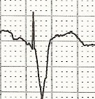
ECG (V1) Sgarbossa criteria, concordant ST depression, ventricular pacing
  • ST elevation = 3.2mm
  • Depth of S wave = 10mm
  • 3.2/10 = 0.32 (32%)
  • ST elevation is 32% of the S wave
  • Criterion 2 is positive
    • (2 points)

Ventricular Pacing and Ventricular Pacing + STEMI

Ventricular Pacing: Ventricular Pacing + Acute STEMI: Criteria:
ECG criteria LBBB without ventricular paced rhythm infarction (V4-V6, aVL, I) ECG Sgarbossa criteria during right ventricular RV pacing and STEMI, concordant ST elevation (V4-V6, aVL, I)
Concordant ST elevation > 1mm
  • 5 points
ECG criteria ventricular paced rhythm without STEMI infarction (V1-V3) ECG Sgarbossa criteria during right ventricular RV pacing and STEMI, disconcordant ST elevation (V1-V3)
Disconcordant ST elevation ≥ 5mm
  • (Or ST elevation > 25% of the depth of the S wave)
  • 2 points
ECG criteria ventricular paced rhythm without STEMI infarction (V1-V3) ECG Sgarbossa criteria during right ventricular RV pacing and STEMI, disconcordant ST depression (V1-V3)
Concordant ST depression > 1mm
  • 3 points

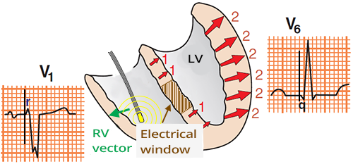
Ventricular Pacing and Old Anteroseptal STEMI

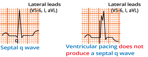
  • Septal Q wave arises from septal activation in lateral leads (V5-V6, I, aVL)
    • The septum is activated from left to right (towards the lateral leads)
  • Ventricular pacing without infarction does not create a Q wave in the lateral leads
    • Because the septum is activated from right to left (towards the lateral leads)
    • The septum is activated by the electrode in the right ventricle
  • If a Q wave is present in the lateral leads (V5-V6, I, aVL) in the context of ventricular pacing
  • Then this Q wave arose due to the electrical window following a septal infarction
Ventricular paced rhythm, and septal Q wave


right ventricular RV pacing rhythm, septal infarction, q wave in lateral ECG leads

Septal Infarction and Ventricular Pacing


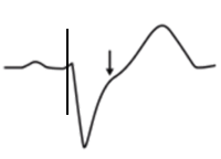
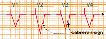
Cabrera Sign and Old Anterior STEMI

  • Old anterior STEMI creates a scar in the myocardium
  • Cabrera Sign
    • Is a notch on the ascending part of the S wave
    • In ventricular pacing
      • Indicates old anterior STEMI
Cabrera sign (shelf-like notch on ascending S wave), old myocardial infarction during right ventricular pacing


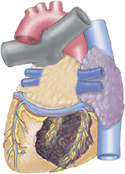
Dual chamber pacing, septal necrosis infarction, old antero-septal infarction
ECG ventricular paced rhythm, old anteroseptal STEMi myocardial infarction, DDD pacing, septal q wave, Cabrera sign

Old Anteroseptal STEMI and Ventricular Pacing


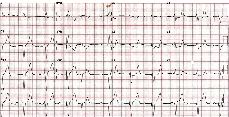
ventricular paced rhythm, posterior infarction, occlusion postero-lateral branch RCA
ECG Positive Sgarbossa criteria in a patient with a ventricular paced rhythm, concordant ST depression

Acute STEMI and Ventricular Pacing

  • Ventricular pacing with LBBB pattern
  • Concordant ST elevation > 1mm (V4-V6, aVL, I)
    • No
  • Disconcordant ST elevation ≥ 5mm (V1-V3)
    • No
  • Concordant ST depression > 1mm (V1-V3)
    • Yes (3 points)
  • The patient has acute STEMI in the territory of ventricular pacing
  • The patient had on coronary angiography
    • Occlusion of a branch of the right coronary artery for the posterior wall
    • Had acute posterior wall STEMI


Sources

  • ECG from Basics to Essentials Step by Step
  • litfl.com
  • ecgwaves.com
  • metealpaslan.com
  • medmastery.com
  • uptodate.com
  • ecgpedia.org
  • wikipedia.org
  • Strong Medicine
  • Understanding Pacemakers


šípka späť

STEMI and Ventricular Pacing

STEMI infarction and ventricular paced rhythm (Sgarbossa criteria)
Ventricular Pacing

  • It is the ability of a pacemaker to stimulate the ventricular myocardium through an electrode
    • The electrode stimulates the myocardium with an electrical impulse (similar to the SA node)
  • The ventricular electrode of the pacemaker is placed in the right ventricle
  • Pacing on the ECG is seen as a pacing artifact (a vertical line, also known as a spike)
  • Ventricular pacing creates a LBBB pattern on the ECG
    • And LBBB masks the ECG signs of infarction

paced ventricular rhythm and acute STEMI infarction

ECG ventricular paced rhythm with LBBB pattern

Sgarbossa Criteria


ECG and Acute STEMI with Ventricular Pacing

  • 1. Concordant ST elevation > 1mm (V4-V6, aVL, I)
    • In leads with a positive QRS complex
    • 5 points
  • 2. Disconcordant ST elevation ≥ 5mm (V1-V3)
    • Or ST elevation > 25% of the depth of the S wave
    • In leads with a negative QRS complex
    • 2 points
  • 3. Concordant ST depression > 1mm (V1-V3)
    • In leads with a negative QRS complex
    • 3 points
  • If the total score is ≥ 3 (Sgarbossa criteria are met)
    • Then the patient has STEMI in the context of
      • Ventricular pacing
      • or LBBB

  • Ventricular pacing on ECG is recognized by
    • stimulus artifact before the QRS complex

ECG Sgarbossa criteria STEMI infarction with ventricular paced rhythm, ST elevation, concordant ST depression


ECG (V1) Sgarbossa criteria, concordant ST depression, ventricular pacing


  • ST elevation = 3.2mm
  • Depth of S wave = 10mm
  • 3.2/10 = 0.32 (32%)
  • ST elevation is 32% of the S wave
  • Criterion 2 is positive
    • (2 points)

Ventricular Pacing and Ventricular Pacing + STEMI

Ventricular Pacing: Ventricular Pacing + Acute STEMI: Criteria:
ECG criteria LBBB without ventricular paced rhythm infarction (V4-V6, aVL, I) ECG Sgarbossa criteria during right ventricular RV pacing and STEMI, concordant ST elevation (V4-V6, aVL, I)
Concordant ST elevation > 1mm
  • 5 points
ECG criteria ventricular paced rhythm without STEMI infarction (V1-V3) ECG Sgarbossa criteria during right ventricular RV pacing and STEMI, disconcordant ST elevation (V1-V3)
Disconcordant ST elevation ≥ 5mm
  • (Or ST elevation > 25% of the depth of the S wave)
  • 2 points
ECG criteria ventricular paced rhythm without STEMI infarction (V1-V3) ECG Sgarbossa criteria during right ventricular RV pacing and STEMI, disconcordant ST depression (V1-V3)
Concordant ST depression > 1mm
  • 3 points

Ventricular Pacing and Old Anteroseptal STEMI

  • Septal Q wave arises from septal activation in lateral leads (V5-V6, I, aVL)
    • The septum is activated from left to right (towards the lateral leads)
  • Ventricular pacing without infarction does not create a Q wave in the lateral leads
    • Because the septum is activated from right to left (towards the lateral leads)
    • The septum is activated by the electrode in the right ventricle
  • If a Q wave is present in the lateral leads (V5-V6, I, aVL) in the context of ventricular pacing
  • Then this Q wave arose due to the electrical window following a septal infarction
Ventricular paced rhythm, and septal Q wave


right ventricular RV pacing rhythm, septal infarction, q wave in lateral ECG leads

Septal Infarction and Ventricular Pacing

Cabrera Sign and Old Anterior STEMI

  • Old anterior STEMI creates a scar in the myocardium
  • Cabrera Sign
    • Is a notch on the ascending part of the S wave
    • In ventricular pacing
      • Indicates old anterior STEMI

Cabrera sign (shelf-like notch on ascending S wave), old myocardial infarction during right ventricular pacing


ECG ventricular paced rhythm, old anteroseptal STEMi myocardial infarction, DDD pacing, septal q wave, Cabrera sign

Old Anteroseptal STEMI and Ventricular Pacing
Dual chamber pacing, septal necrosis infarction, old antero-septal infarction


ECG Positive Sgarbossa criteria in a patient with a ventricular paced rhythm, concordant ST depression

Acute STEMI and Ventricular Pacing

  • Ventricular pacing with LBBB pattern
  • Concordant ST elevation > 1mm (V4-V6, aVL, I)
    • No
  • Disconcordant ST elevation ≥ 5mm (V1-V3)
    • No
  • Concordant ST depression > 1mm (V1-V3)
    • Yes (3 points)
  • The patient has acute STEMI in the territory of ventricular pacing
  • The patient had on coronary angiography
    • Occlusion of a branch of the right coronary artery for the posterior wall
    • Had acute posterior wall STEMI
ventricular paced rhythm, posterior infarction, occlusion postero-lateral branch RCA


Sources

  • ECG from Basics to Essentials Step by Step
  • litfl.com
  • ecgwaves.com
  • metealpaslan.com
  • medmastery.com
  • uptodate.com
  • ecgpedia.org
  • wikipedia.org
  • Strong Medicine
  • Understanding Pacemakers