|
|
ECGbook.com Making Medical Education Free for All |
Upload ECG for Interpretation |
|
|
ECGbook.com Making Medical Education Free for All |
Upload ECG for Interpretation |
|
|
ECGbook.com Making Medical Education Free for All |
Home /
Supraventricular tachycardia (SVT), Narrow complex tachycardia


Basic Classification of Tachycardias
Narrow Complex Tachycardia

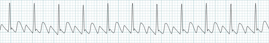
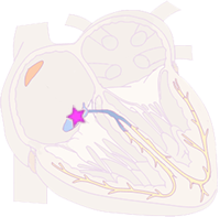
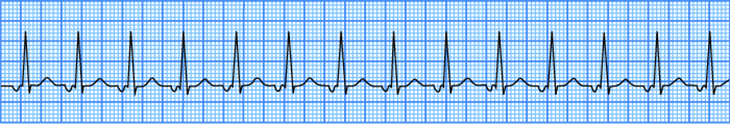
Sinus Tachycardia

Inappropriate Sinus Tachycardia

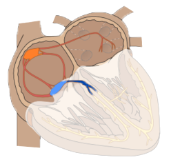
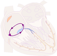
Sinoatrial Nodal Reentry Tachycardia


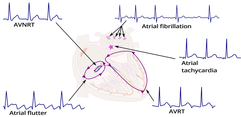
Atrial Tachycardia
Intra-Atrial Reentrant Tachycardia

Multifocal Atrial Tachycardia
Atrial Fibrillation

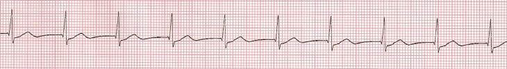
Atrial Flutter
Junctional Tachycardia

Nonparoxysmal Junctional Tachycardia
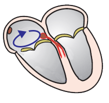
AV Nodal Reentry Tachycardia (AVNRT)

AV Reentry Tachycardia (AVRT)
Permanent Junctional Reciprocal Tachycardia (PJRT)
Sources
Home /
Supraventricular tachycardia (SVT), Narrow complex tachycardia
Supraventricular Impulse
|
|
Narrow QRS Complex
|
|
Basic Classification of Tachycardias
|
Narrow Complex Tachycardia
|
|
|
|
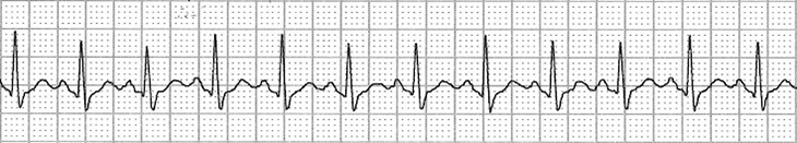
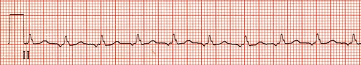
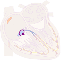
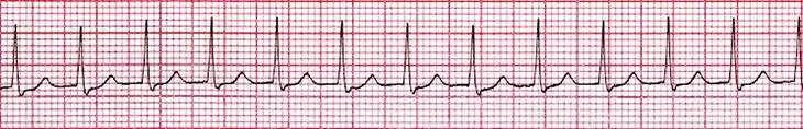
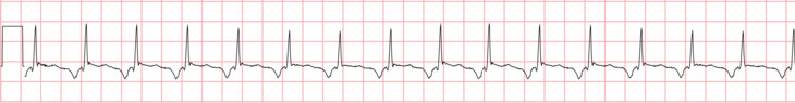
Sinus Tachycardia
|
|
|
Inappropriate Sinus Tachycardia
|
|
|
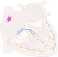
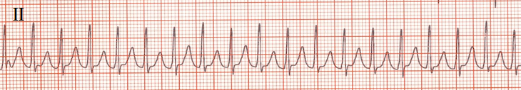
Sinoatrial Nodal Reentry Tachycardia
|
|
|
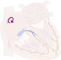
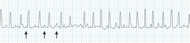
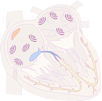
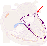
Atrial Tachycardia
|
|
|
Intra-Atrial Reentrant Tachycardia
|
|
|
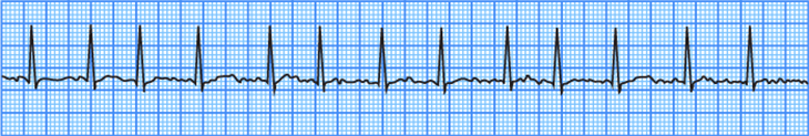
Multifocal Atrial Tachycardia
|
|
|
Atrial Fibrillation
|
|
|
Atrial Flutter
|
|
|
Junctional Tachycardia
|
|
|
Nonparoxysmal Junctional Tachycardia
|
|
|
AV Nodal Reentry Tachycardia (AVNRT)
|
|
|
AV Reentry Tachycardia (AVRT)
|
|
|
Permanent Junctional Reciprocal Tachycardia (PJRT)
|
Sources