ECGbook.com

Making Medical Education Free for All

ECGbook.com

Making Medical Education Free for All

Home /

Ventricular Rhythm

Ventricular rhythm, Idioventricular escape rhythm

Heart Rhythm

heart rhythm mechanism, sinus rhythm, junctional rhythm, ventricular rhythm

Basic Heart Rhythms

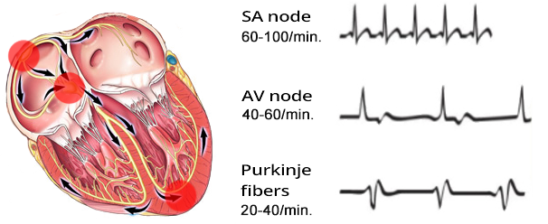
  • Heart rhythm is always determined by the site (sites) that generate impulses with the highest frequency (overdrive suppression)
  • There are 3 basic heart rhythms
  • Sinus Rhythm
    • It is the primary pacemaker
    • Generates impulses with a frequency of: 60 - 100/min.
  • Junctional Rhythm
    • It is the secondary pacemaker
    • Generates impulses with a frequency of: 40 - 60/min.
  • Ventricular Rhythm
    • It is the tertiary pacemaker
    • Generates impulses with a frequency of: 20 - 40/min.
    • Activates when:
      • Impulses from the supraventricular area (SA node and AV junction) cease to reach the ventricles
      • The ventricles outpace the supraventricular areas in generating impulses

Ventricular Rhythm

Ventricular, idioventricular escape rhythm

Ventricular Rhythm - Most Common Causes


QRS Complex Width and Cardiac Rhythms

sinus rhythm
ECG sinus rhythm, narrow QRS complexes

Sinus Rhythm


junctional rhythm
ECG junctional rhythm, narrow QRS complexes

Junctional Rhythm


ventricular rhythm
ECG ventricular escape rhythm

Ventricular Rhythm


ECG and Ventricular Rhythm

ventricular ectopic focus, and ventricular escape rhythm

ECG ventricular rhythm, broad qrs complexes

Ventricular Rhythm


Ectopic Focus Localization


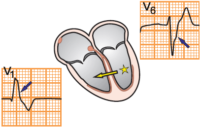
ecg V1, V6 ectopic ventricular focus, lbbb pattern

Ectopic Focus in the Right Ventricle

  • Negative QRS (V1)
    • Vector points away from lead V1
  • Positive QRS (V6)
    • Vector points toward lead V6


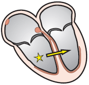
ecg V1, V6 ectopic ventricular focus, rbbb pattern

Ectopic Focus in the Left Ventricle

  • Positive QRS (V1)
    • Vector points toward lead V1
  • Negative QRS (V6)
    • Vector points away from lead V6

Frequency and Ventricular Rhythm


ECG ventricular escape rhythm, SA block 3rd degree

Ventricular Rhythm

  • Frequency: 27/min.
    • Ventricular rhythm has a frequency of 20-40/min.
  • Wide QRS complexes (> 0.12s)
  • P waves are absent
  • Often, a secondary pacemaker is activated - junctional rhythm
    • In this case, the ventricular rhythm - tertiary pacemaker was activated


ECG accelerated idio-ventricular rhythm

Accelerated Ventricular Rhythm


ECG ventricular tachycardia, 160bpm, broad QRS complexes

Ventricular Tachycardia

  • Frequency: 160/min.
  • Wide QRS complexes (> 0.12s)
  • P waves are absent
  • Patients with ventricular tachycardia are often hemodynamically unstable, with a risk of ventricular fibrillation
    • The heart stops functioning as a pump
    • Requires urgent treatment (medication or defibrillation)


idio-ventricular ectopic focus, and lbbb pattern
ECG idio-ventricular escape rhythm, and sinus pause

Ventricular Rhythm and Sinus Pause


ventricular ectopic focus, RBBB pattern V1, V6
ecg idio-venricular escape rhythm, complete 3rd degree av block, av dissociation

Ventricular Rhythm and AV Block III Degree
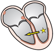
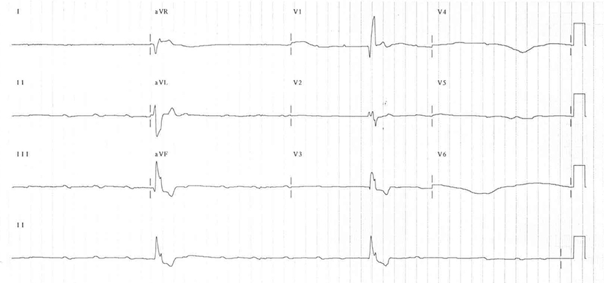
  • Frequency 27/min.
  • Wide QRS complexes (> 0.12s)
  • Heart rate is regular (RR interval is constant)
  • The ectopic focus is in the left ventricle (see image of RBBB)
    • Positive QRS (V1)
    • Negative QRS (V6)
  • The ectopic focus is in the left ventricle in the area of the left anterior fascicle
  • On the ECG, there is an AV Block III Degree
    • There is electrical isolation between the atria and ventricles
      • The AV junction is interrupted
    • Frequency of P waves is 65/min. (Continuous II lead)
      • Impulses from the SA node are blocked at the AV junction
    • AV Dissociation
      • There are 2 independent pacemakers in the heart: (that generate impulses)
        • SA node
        • Ectopic ventricular focus
      • P waves and QRS complexes are completely independent of each other


ventricular ectopic focus, RBBB pattern
ECG idio-ventricular rhythm, and 3rd degree av complete block, av dissociation, broad qrs

Ventricular Rhythm and AV Block III Degree


Sources
  • ECG from Basics to Essentials Step by Step
  • litfl.com
  • ecgwaves.com
  • metealpaslan.com
  • medmastery.com
  • uptodate.com
  • ecgpedia.org
  • wikipedia.org
  • Strong Medicine
  • Understanding Pacemakers


šípka späť

Ventricular Rhythm

Ventricular rhythm, Idioventricular escape rhythm

Heart Rhythm

heart rhythm mechanism, sinus rhythm, junctional rhythm, ventricular rhythm

Basic Heart Rhythms

  • Heart rhythm is always determined by the site (sites) that generate impulses with the highest frequency (overdrive suppression)
  • There are 3 basic heart rhythms
  • Sinus Rhythm
    • It is the primary pacemaker
    • Generates impulses with a frequency of: 60 - 100/min.
  • Junctional Rhythm
    • It is the secondary pacemaker
    • Generates impulses with a frequency of: 40 - 60/min.
  • Ventricular Rhythm
    • It is the tertiary pacemaker
    • Generates impulses with a frequency of: 20 - 40/min.
    • Activates when:
      • Impulses from the supraventricular area (SA node and AV junction) cease to reach the ventricles
      • The ventricles outpace the supraventricular areas in generating impulses

Ventricular Rhythm

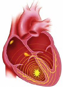
  • Impulses originate in the ventricles
  • Each impulse then activates the ventricles through the myocardium
    • Impulses spread slowly through the myocardium
    • Therefore, QRS complexes will be wide (>0.12s)
  • Ventricular rhythm is very rare, it activates
    • In case of SA node and AV junction failure
  • Ventricular rhythm has a frequency of 20-40/min.
    • Sometimes referred to as
      • Idioventricular rhythm
      • Ventricular escape rhythm

Ventricular, idioventricular escape rhythm

Ventricular Rhythm - Most Common Causes


QRS Complex Width and Cardiac Rhythms

sinus rhythm ECG sinus rhythm, narrow QRS complexes

Sinus Rhythm

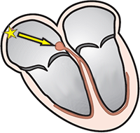
  • SA node generates impulses with a frequency: 60-100/min.
  • Atrial vector directs towards the AV node (to Lead II)
    • In Lead II, a positive P wave forms
  • Then the ventricles are activated
  • First, the atria are activated (P wave) and then the ventricles (QRS)
  • On the ECG, it is a sinus rhythm


junctional rhythm ECG junctional rhythm, narrow QRS complexes

Junctional Rhythm


ventricular rhythm ECG ventricular escape rhythm

Ventricular Rhythm

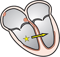
  • Impulses are generated in the ventricles with a frequency of: 20-40/min.
  • The impulse spreads from the ventricles:
    • Slowly through the ventricular myocardium
    • The impulse does not activate the ventricles via the conduction system
  • A wide QRS complex (>0.12s) forms
    • Because the impulse spreads more slowly through the myocardium compared to the conduction system
  • The impulse may sometimes pass through the AV junction and retrogradely activate the atria

ECG and Ventricular Rhythm


ventricular ectopic focus, and ventricular escape rhythm


ECG ventricular rhythm, broad qrs complexes

Ventricular Rhythm


Ectopic Focus Localization


ecg V1, V6 ectopic ventricular focus, lbbb pattern

Ectopic Focus in the Right Ventricle

  • Negative QRS (V1)
    • Vector points away from lead V1
  • Positive QRS (V6)
    • Vector points toward lead V6
ecg V1, V6 ectopic ventricular focus, rbbb pattern

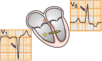
Ectopic Focus in the Left Ventricle

  • Positive QRS (V1)
    • Vector points toward lead V1
  • Negative QRS (V6)
    • Vector points away from lead V6

Frequency and Ventricular Rhythm


ECG ventricular escape rhythm, SA block 3rd degree

Ventricular Rhythm

  • Frequency: 27/min.
    • Ventricular rhythm has a frequency of 20-40/min.
  • Wide QRS complexes (> 0.12s)
  • P waves are absent
  • Often, a secondary pacemaker is activated - junctional rhythm
    • In this case, the ventricular rhythm - tertiary pacemaker was activated


ECG accelerated idio-ventricular rhythm

Accelerated Ventricular Rhythm


ECG ventricular tachycardia, 160bpm, broad QRS complexes

Ventricular Tachycardia

  • Frequency: 160/min.
  • Wide QRS complexes (> 0.12s)
  • P waves are absent
  • Patients with ventricular tachycardia are often hemodynamically unstable, with a risk of ventricular fibrillation
    • The heart stops functioning as a pump
    • Requires urgent treatment (medication or defibrillation)


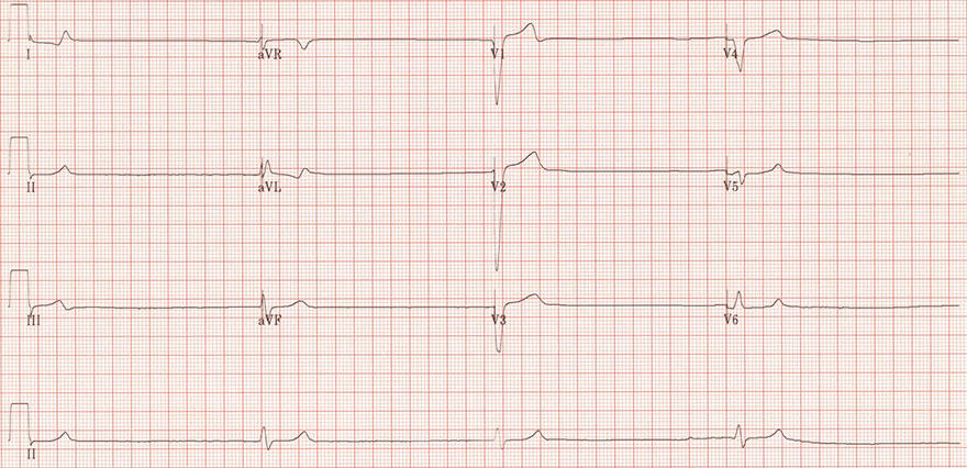
ECG idio-ventricular escape rhythm, and sinus pause

Ventricular Rhythm and Sinus Pause

  • Frequency 25/min.
  • Wide QRS complexes (> 0.12s)
  • Heart rate is regular (RR interval is constant)
  • The ectopic focus is in the right ventricle (see image of LBBB)
    • Negative QRS (V1)
    • Positive QRS (V6)
  • On the ECG, there is a sinus pause
    • At the end of the continuous II lead, we see a P wave
    • Due to increased parasympathetic tone, the junctional rhythm did not activate
idio-ventricular ectopic focus, and lbbb pattern


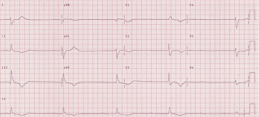
ecg idio-venricular escape rhythm, complete 3rd degree av block, av dissociation

Ventricular Rhythm and AV Block III Degree

  • Frequency 27/min.
  • Wide QRS complexes (> 0.12s)
  • Heart rate is regular (RR interval is constant)
  • The ectopic focus is in the left ventricle (see image of RBBB)
    • Positive QRS (V1)
    • Negative QRS (V6)
  • The ectopic focus is in the left ventricle in the area of the left anterior fascicle
  • On the ECG, there is an AV Block III Degree
    • There is electrical isolation between the atria and ventricles
      • The AV junction is interrupted
    • Frequency of P waves is 65/min. (Continuous II lead)
      • Impulses from the SA node are blocked at the AV junction
    • AV Dissociation
      • There are 2 independent pacemakers in the heart: (that generate impulses)
        • SA node
        • Ectopic ventricular focus
      • P waves and QRS complexes are completely independent of each other
ventricular ectopic focus, RBBB pattern V1, V6


ECG idio-ventricular rhythm, and 3rd degree av complete block, av dissociation, broad qrs

Ventricular Rhythm and AV Block III Degree

  • Frequency 15/min.
  • Wide QRS complexes (> 0.12s)
  • Heart rate is regular (RR interval is constant)
  • The ectopic focus is in the left ventricle (see image of RBBB)
    • Positive QRS (V1)
  • On the ECG, there is an AV Block III Degree
    • The atria and ventricles are electrically isolated from each other
      • The AV junction is interrupted
    • Frequency of P waves is 100/min. (Continuous II lead)
      • Impulses from the SA node are blocked at the AV junction
  • AV Dissociation
    • There are 2 independent pacemakers in the heart: (that generate impulses)
      • SA node
      • Ectopic ventricular focus
    • P waves and QRS complexes are completely independent of each other
ventricular ectopic focus, RBBB pattern


Sources

  • ECG from Basics to Essentials Step by Step
  • litfl.com
  • ecgwaves.com
  • metealpaslan.com
  • medmastery.com
  • uptodate.com
  • ecgpedia.org
  • wikipedia.org
  • Strong Medicine
  • Understanding Pacemakers