ECGbook.com

Making Medical Education Free for All

ECGbook.com

Making Medical Education Free for All

Home /

aVR Algorithm (Vereckei)

Vereckei aVR algorithm (ventricular tachycardia vs. SVT with aberrant conduction)
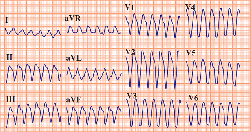
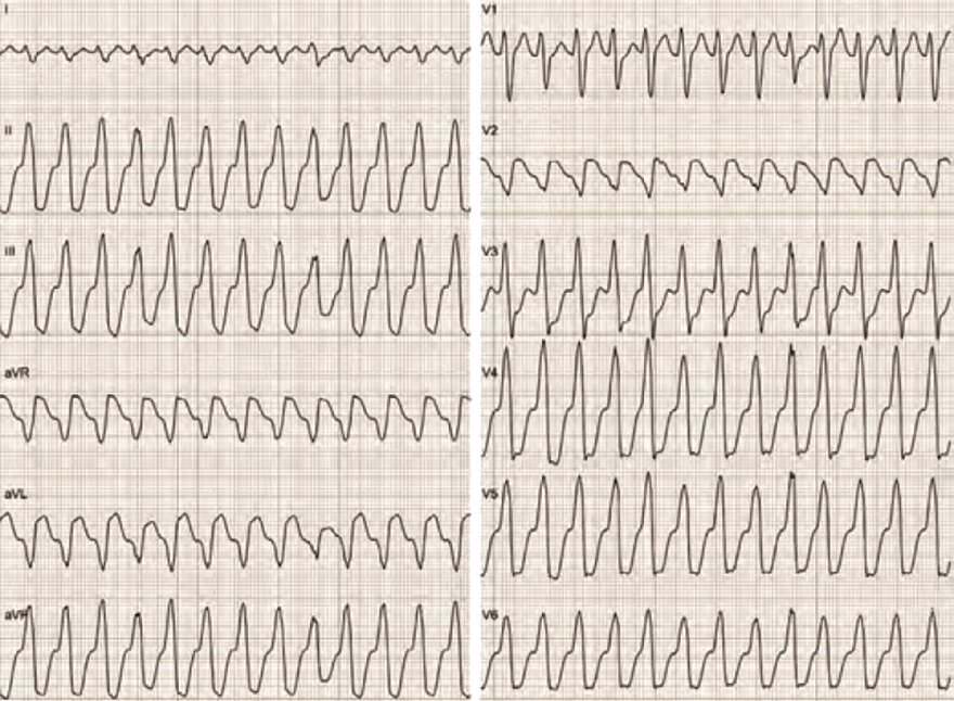
Ventricular Tachycardia


Ventricular tachycardia, aVR algorithm - Vereckei

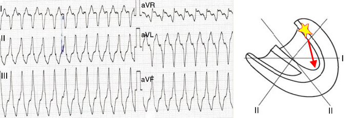
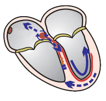
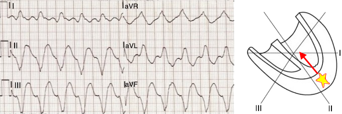
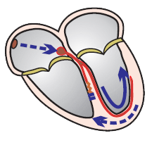
Ectopic Focus in Ventricular Tachycardia

  • Most commonly located in the lower part of the heart, in the apex
  • The resulting ventricular vector thus points upwards towards the aVR lead (from lead II)
    • This is the main diagnostic principle of the aVR algorithm

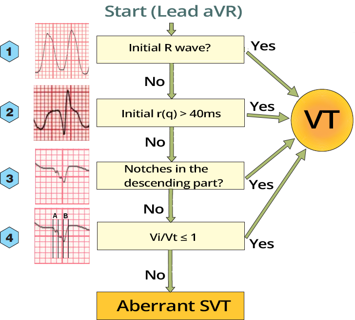
aVR Algorithm


aVR algorithm: Difference between VT vs SVT with aberrancy on ECG

Wide-Complex Tachycardia


aVR Algorithm in Clinical Practice

  • Sensitivity: 96%, Specificity: 99%
  • aVR Algorithm
    • Sequentially evaluates 4 ECG criteria (only in the aVR lead); if none apply, it is Aberrant SVT
  • Differentiates wide-complex tachycardia: distinguishes VT from Aberrant SVT


DDx wide complex tachycardia (aVR algorithm - vereckei), initial R wave, Initial r(q) 40ms, Notch on downstroke QRS, Vi/Vt ratio

aVR Algorithm (Evaluates only the aVR Lead)


aVR Algorithm - Limitations

  • The aVR algorithm is very fast and simple, as it evaluates only the aVR lead
  • It is based on the principle that an ectopic focus in ventricular tachycardia (VT) is most commonly located in the lower part of the heart
    • In such cases, the resulting vector points toward the aVR lead and VT is diagnosed immediately in the 1st step of the algorithm

aVR algorithm - Vereckei, Ventricular tachycardia, aVR algorithm - Vereckei, apex localized ectopic ventricular focus

Wide-Complex Tachycardia


aVR algorithm - Vereckei, Ventricular tachycardia, base localised ectopic focus, negative QRS in aVR lead

Wide-Complex Tachycardia


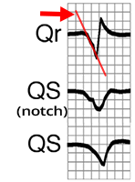
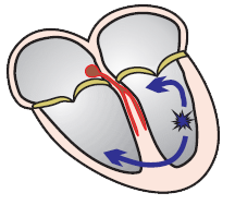
Depolarization in VT

ECG Qr, QS, Lead aVR, slow depolarization from ventricular ectopic focus (ventricular tachycardia), Notch in descending limb, broad q(r) wave aVR algorithm - Andras Vereckei, initial slow depolarization rate
  • In VT, the ventricles are depolarized from a ventricular ectopic focus
  • Depolarization through the myocardium is slow
    • Therefore, the beginning of the QRS is less steep
      • Compared to depolarization through the conduction system
  • Notch most commonly occurs in a structurally altered heart
  • From this fact follow
    • Steps 2, 3, and 4 of the aVR algorithm

Depolarization in Aberrant SVT

ECG fast depolarization, aVR lead (Vereckei), supraventricular tachycardia with aberrant conduction aVR algorithm - Andras Vereckei, Wide complex tachycardia, SVT with aberrant conduction due to bundle branch block, RBBB, LBBB

Vi/Vt ≤ 1

  • It is the 4th step of the aVR algorithm
  • Vi is the distance (mV) that the impulse travels at the beginning of depolarization (at the beginning of QRS) in 40ms (in 1 small square)
  • Vt is the distance (mV) that the impulse travels at the end of depolarization (at the end of QRS) in 40ms (in 1 small square)
  • If the Vi/Vt ratio ≤ 1, it indicates ventricular tachycardia
    • The formula is based on the principle that in VT, the initial part of the QRS is less steep compared to SVT


Vereckei aVR algorithm, vi vt ratio, ddx wide complex tachycardia
  • Step 4 of the aVR algorithm in practice is impractical
  • It is best to mark the start and end of the QRS with a ruler, and then measure 40ms in aVR (1 small square)
Lead aVR Vereckei, Vi/Vt, ventricular tachycardia - VT diagnosed

Wide-Complex Tachycardia

  • Vi = 1.5mm (A: height of the curve in 40ms is 1mm)
  • Vt = 6mm (B: height of the curve in 40ms is 3mm)
  • Vi/Vt (1.5/6 = 0.25) ≤ 1, indicating ventricular tachycardia


Lead aVR Vereckei, Vi/Vt, supraventricular tachycardia - SVT diagnosed

Wide-Complex Tachycardia

  • Vi = 4mm (A: height of the curve in 40ms is 1mm)
  • Vt = 2mm (B: height of the curve in 40ms is 3mm)
  • Vi/Vt (4/2 = 2) ≤ 1, indicating Aberrant SVT


aVR algorithm - Andras Vereckei, Wide complex tachycardia
ECG differential diagnosis (DDx) wide compelex tachycardia, aVR Vereckei algorithm, initial R wave (aVR) - ventricular tachycardia

Wide-Complex Tachycardia


aVR algorithm - Andras Vereckei, Wide complex tachycardia
ECG DDx wide complex tachycardia, aVR Vereckei algorithm, Initial R wave, Initial r(q) 40ms, Ventricular tachycardia

Wide-Complex Tachycardia


AVNRT with RBBB, anterograde ventricle depolarization, retrograde atria depolarization
ECG differential diagnosis (DDx) wide compelex tachycardia, aVR Vereckei algorithm, Initial R - NO, Inital r(q) 40ms - NO, Notch - NO, Vi Vt - NO, SVT with RBBB diagnosed

Wide-Complex Tachycardia


Sources

  • ECG from Basics to Essentials Step by Step
  • litfl.com
  • ecgwaves.com
  • metealpaslan.com
  • medmastery.com
  • uptodate.com
  • ecgpedia.org
  • wikipedia.org
  • Strong Medicine
  • Understanding Pacemakers


šípka späť

aVR Algorithm (Vereckei)

Vereckei aVR algorithm (ventricular tachycardia vs. SVT with aberrant conduction)

Ventricular Tachycardia


Ventricular tachycardia, aVR algorithm - Vereckei

Ectopic Focus in Ventricular Tachycardia

  • Most commonly located in the lower part of the heart, in the apex
  • The resulting ventricular vector thus points upwards towards the aVR lead (from lead II)
    • This is the main diagnostic principle of the aVR algorithm

aVR Algorithm


aVR algorithm - Andras Vereckei, Wide complex tachycardia aVR algorithm - Andras Vereckei, Wide complex tachycardia, SVT with aberrant conduction due to bundle branch block, RBBB, LBBB aVR algorithm - Andras Vereckei, Wide complex tachycardia, Orthodromic AV, SVT with aberrant conduction due to the Wolff-Parkinson-White (WPW) syndrome
arrows
aVR algorithm - Andras Vereckei, Differential Diagnosis (DDx) of Wide-Complex Tachycardia, ventricular tachycardia

Wide-Complex Tachycardia


aVR Algorithm in Clinical Practice

  • Sensitivity: 96%, Specificity: 99%
  • aVR Algorithm
    • Sequentially evaluates 4 ECG criteria (only in the aVR lead); if none apply, it is Aberrant SVT
  • Differentiates wide-complex tachycardia: distinguishes VT from Aberrant SVT


DDx wide complex tachycardia (aVR algorithm - vereckei), initial R wave, Initial r(q) 40ms, Notch on downstroke QRS, Vi/Vt ratio

aVR Algorithm (Evaluates only the aVR Lead)


aVR Algorithm - Limitations

  • The aVR algorithm is very fast and simple, as it evaluates only the aVR lead
  • It is based on the principle that an ectopic focus in ventricular tachycardia (VT) is most commonly located in the lower part of the heart
    • In such cases, the resulting vector points toward the aVR lead and VT is diagnosed immediately in the 1st step of the algorithm

aVR algorithm - Vereckei, Ventricular tachycardia, aVR algorithm - Vereckei, apex localized ectopic ventricular focus

Wide-Complex Tachycardia


aVR algorithm - Vereckei, Ventricular tachycardia, base localised ectopic focus, negative QRS in aVR lead

Wide-Complex Tachycardia


Depolarization in VT

ECG Qr, QS, Lead aVR, slow depolarization from ventricular ectopic focus (ventricular tachycardia), Notch in descending limb, broad q(r) wave aVR algorithm - Andras Vereckei, initial slow depolarization rate
  • In VT, the ventricles are depolarized from a ventricular ectopic focus
  • Depolarization through the myocardium is slow
    • Therefore, the beginning of the QRS is less steep
      • Compared to depolarization through the conduction system
  • Notch most commonly occurs in a structurally altered heart
  • From this fact follow
    • Steps 2, 3, and 4 of the aVR algorithm

Depolarization in Aberrant SVT

ECG fast depolarization, aVR lead (Vereckei), supraventricular tachycardia with aberrant conduction aVR algorithm - Andras Vereckei, Wide complex tachycardia, SVT with aberrant conduction due to bundle branch block, RBBB, LBBB
  • In SVT, the ventricles are depolarized
    • Through the conduction system, not through the myocardium
  • Depolarization through the conduction system is fast
    • Therefore, the beginning of the QRS is steeper
      • Compared to depolarization through the myocardium in VT
  • The beginning of the QRS is steep even in SVT with bundle branch block
    • Because one ventricle is depolarized quickly
      • Through the intact bundle branch

Vi/Vt ≤ 1

  • It is the 4th step of the aVR algorithm
  • Vi is the distance (mV) that the impulse travels at the beginning of depolarization (at the beginning of QRS) in 40ms (in 1 small square)
  • Vt is the distance (mV) that the impulse travels at the end of depolarization (at the end of QRS) in 40ms (in 1 small square)
  • If the Vi/Vt ratio ≤ 1, it indicates ventricular tachycardia
    • The formula is based on the principle that in VT, the initial part of the QRS is less steep compared to SVT


Vereckei aVR algorithm, vi vt ratio, ddx wide complex tachycardia
  • Vi = 1mm (A: the height of the curve in 40ms is 1mm)
  • Vt = 3mm (B: the height of the curve in 40ms is 3mm)
  • Vi/Vt (1/3 = 0.33) ≤ 1, indicating ventricular tachycardia

  • According to the given ECG curve, using the aVR algorithm
    • You would diagnose VT already in the 2nd step - initial Q width is > 40ms (80ms)

  • Step 4 of the aVR algorithm in practice is impractical
  • It is best to mark the start and end of the QRS with a ruler, and then measure 40ms in aVR (1 small square)

Lead aVR Vereckei, Vi/Vt, ventricular tachycardia - VT diagnosed

Wide-Complex Tachycardia

  • Vi = 1.5mm (A: height of the curve in 40ms is 1mm)
  • Vt = 6mm (B: height of the curve in 40ms is 3mm)
  • Vi/Vt (1.5/6 = 0.25) ≤ 1, indicating ventricular tachycardia
Lead aVR Vereckei, Vi/Vt, supraventricular tachycardia - SVT diagnosed

Wide-Complex Tachycardia

  • Vi = 4mm (A: height of the curve in 40ms is 1mm)
  • Vt = 2mm (B: height of the curve in 40ms is 3mm)
  • Vi/Vt (4/2 = 2) ≤ 1, indicating Aberrant SVT


ECG differential diagnosis (DDx) wide compelex tachycardia, aVR Vereckei algorithm, initial R wave (aVR) - ventricular tachycardia

Wide-Complex Tachycardia

aVR algorithm - Andras Vereckei, Wide complex tachycardia


ECG DDx wide complex tachycardia, aVR Vereckei algorithm, Initial R wave, Initial r(q) 40ms, Ventricular tachycardia

Wide-Complex Tachycardia

aVR algorithm - Andras Vereckei, Wide complex tachycardia


ECG differential diagnosis (DDx) wide compelex tachycardia, aVR Vereckei algorithm, Initial R - NO, Inital r(q) 40ms - NO, Notch - NO, Vi Vt - NO, SVT with RBBB diagnosed

Wide-Complex Tachycardia

  • QRS Duration: 0.14s
  • Heart Rate 212/min.
  • aVR Algorithm
    • Condition 1: Is there an initial R (aVR) - NO
    • Condition 2: Initial r(q) > 40ms - NO
    • Condition 3: Notches on the descending limb? - NO
    • Condition 4: Vi/Vt ≤ 1 - NO
  • It is SVT with RBBB (Specifically, AVNRT with RBBB)
    • Typical Image of RBBB (In V1, rsR' - right bunny ear is larger)
AVNRT with RBBB, anterograde ventricle depolarization, retrograde atria depolarization


Sources

  • ECG from Basics to Essentials Step by Step
  • litfl.com
  • ecgwaves.com
  • metealpaslan.com
  • medmastery.com
  • uptodate.com
  • ecgpedia.org
  • wikipedia.org
  • Strong Medicine
  • Understanding Pacemakers