|
|
ECGbook.com Making Medical Education Free for All |
Upload ECG for Interpretation |
|
|
ECGbook.com Making Medical Education Free for All |
Upload ECG for Interpretation |
|
|
ECGbook.com Making Medical Education Free for All |
Home /
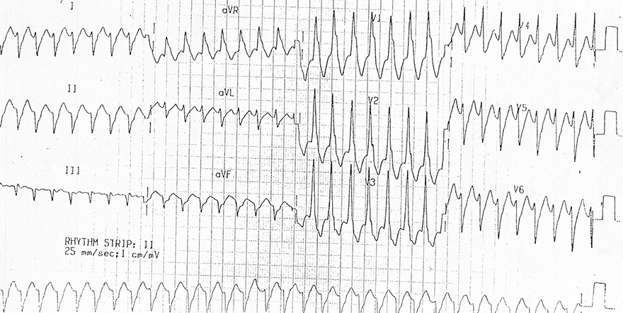
Localisation of the origin of a ventricular tachycardia
| ECG finding: | Localization: |
| QRS Morphology: | |
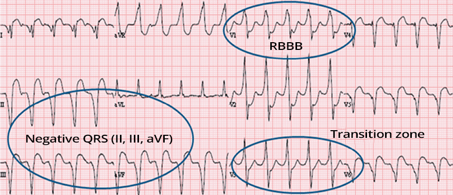
| Shape of LBBB | Left ventricular septum, Right ventricle, Bundle branch reentry VT |
| Shape of RBBB | Left ventricle outside the septum |
| Cardiac Axis: | |
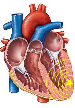
| Superior | Inferior wall |
| Inferior | Upper septum, Upper lateral wall |
| Rightward | Left lateral wall, Apex |
| Transition Zone: | |
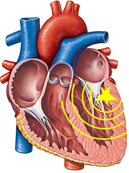
| Transition zone ≤ V3 | Base of the heart |
| Transition zone ≥ V4 | Apex of the heart |
| Precordial Concordance: | |
| Positive | Base of the heart |
| Negative | Apex of the heart |
Ventricular Tachycardia
| ECG Criteria: | Definition: |
| Pseudo delta wave ≥ 35ms | From the beginning of the QRS to the end of the pseudo delta wave In any precordial lead |
| Prolonged R wave peak time ≥ 85ms | From the beginning of the QRS to the peak of the R wave In lead V2 |
| RS interval ≥ 120ms | From the beginning of the QRS to the peak of the S wave In any lead |
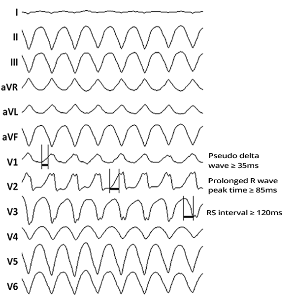
Ventricular Tachycardia
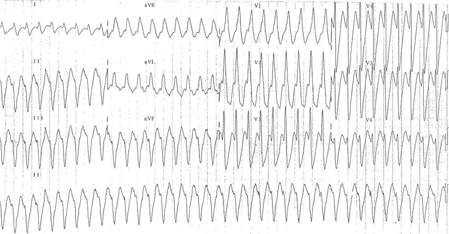
Ventricular Tachycardia
Sources
Home /
Localisation of the origin of a ventricular tachycardia
Ventricular Tachycardia
|
|
| ECG finding: | Localization: |
| QRS Morphology: | |
| Shape of LBBB | Left ventricular septum, Right ventricle, Bundle branch reentry VT |
| Shape of RBBB | Left ventricle outside the septum |
| Cardiac Axis: | |
| Superior | Inferior wall |
| Inferior | Upper septum, Upper lateral wall |
| Rightward | Left lateral wall, Apex |
| Transition Zone: | |
| Transition zone ≤ V3 | Base of the heart |
| Transition zone ≥ V4 | Apex of the heart |
| Precordial Concordance: | |
| Positive | Base of the heart |
| Negative | Apex of the heart |
|
Ventricular Tachycardia
|
|
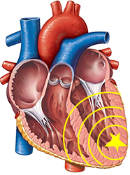
Epicardial VT
|
|
| ECG Criteria: | Definition: |
| Pseudo delta wave ≥ 35ms | From the beginning of the QRS to the end of the pseudo delta wave In any precordial lead |
| Prolonged R wave peak time ≥ 85ms | From the beginning of the QRS to the peak of the R wave In lead V2 |
| RS interval ≥ 120ms | From the beginning of the QRS to the peak of the S wave In any lead |
|
Ventricular Tachycardia
|
|
|
Ventricular Tachycardia
|
|
Sources